5 minutes on…The Larynx.
The Larynx. A total nightmare to learn about. Still, where would we be without her? Yapping a lot less, that’s for sure.
Gross Overview
The larynx is located in the anterior neck
Spans the vertebral levels of C3 to C6
People often confuse the pharynx and larynx
The pharynx leads to the oesophagus, whereas the larynx leads to the trachea
So whilst both air and food passes through the pharynx, only air must pass through the larynx, otherwise food would enter your lungs
Function of the Larynx
Phonation - voice production
Airway protection - prevention of food being aspirated
Breathing - allows air to pass to the lungs
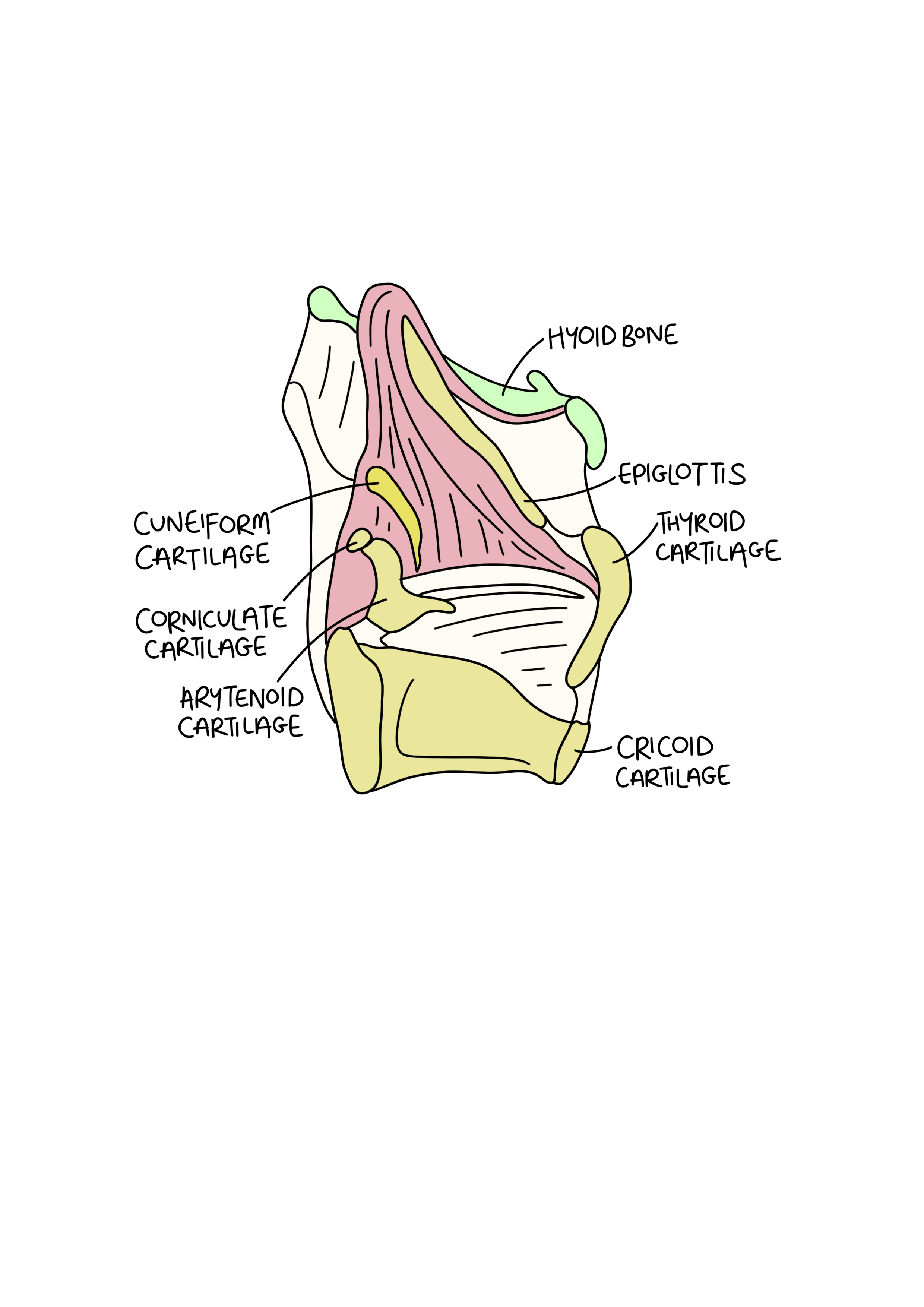
The Laryngeal Cartilages
When covering regions of the body, we often cover bones, or osteology, first. However, the larynx has a cartilaginous skeleton - so let’s cover that in 5 minutes instead. Once you have an understanding of the cartilages it will be much easier to build in the knowledge about the ligaments, muscles, nerves and their locations.
The cartilages can be divided into unpaired and paired cartilages.
Unpaired
Epiglottic cartilage
Thyroid cartilage
Cricoid cartilage
Paired
Arytenoid cartilages
Corniculate cartilages
Cuneiform cartilages
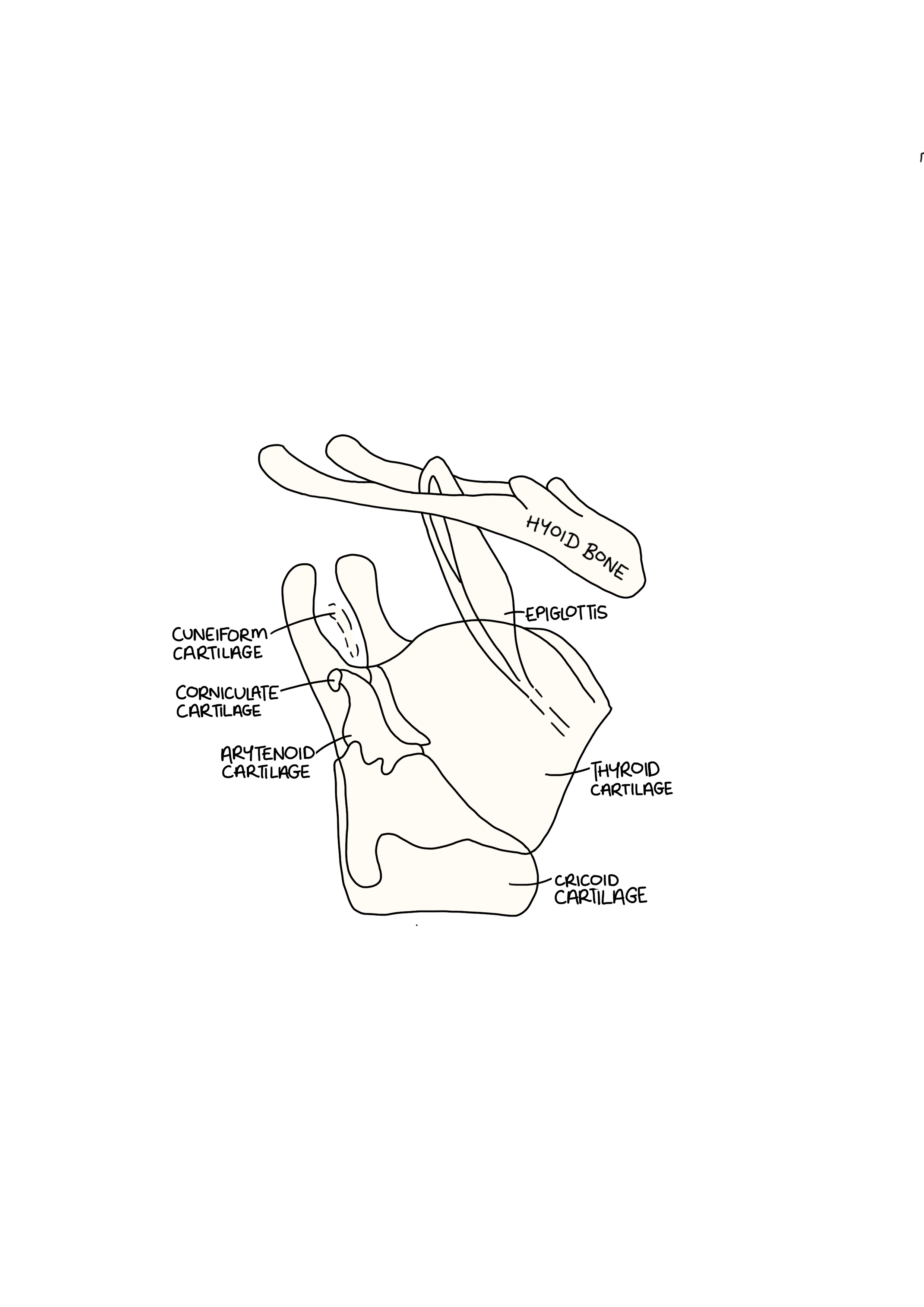
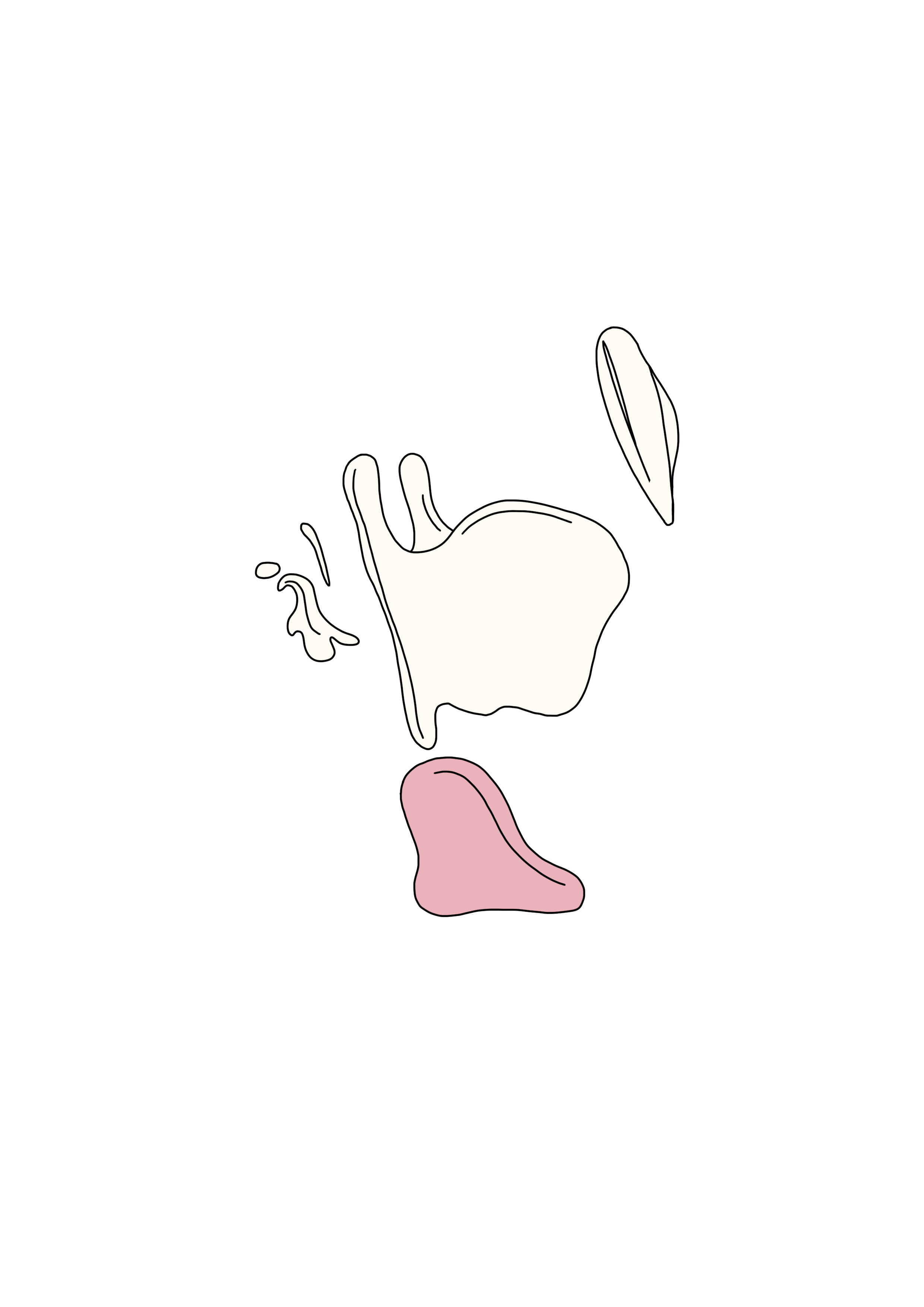
Here they all are together as a unit (with the hyoid bone also featured, which is of course, not a cartilage).
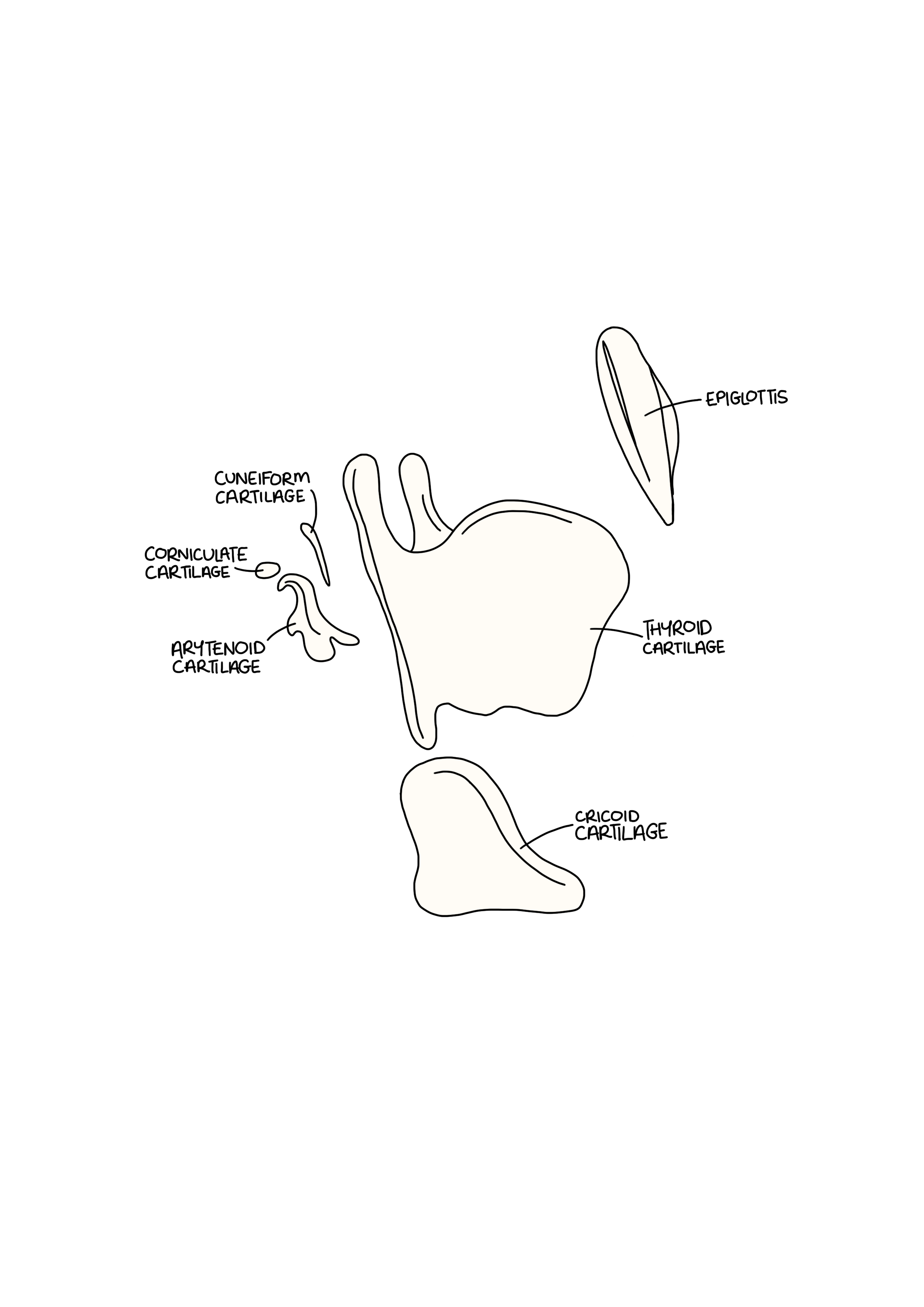
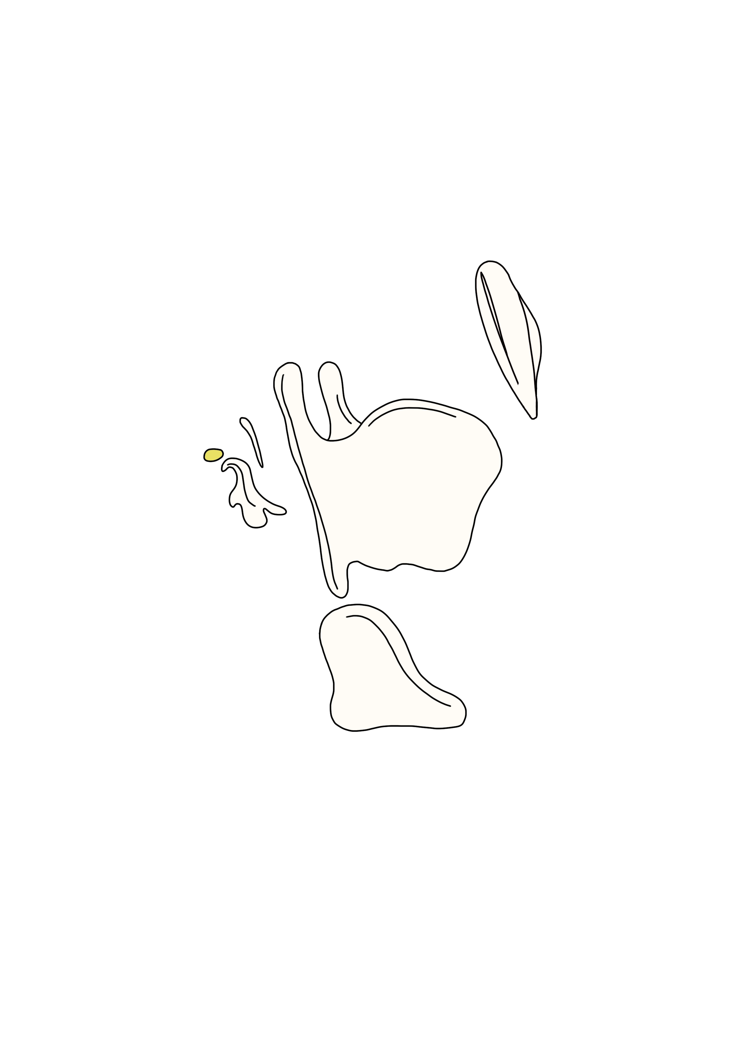
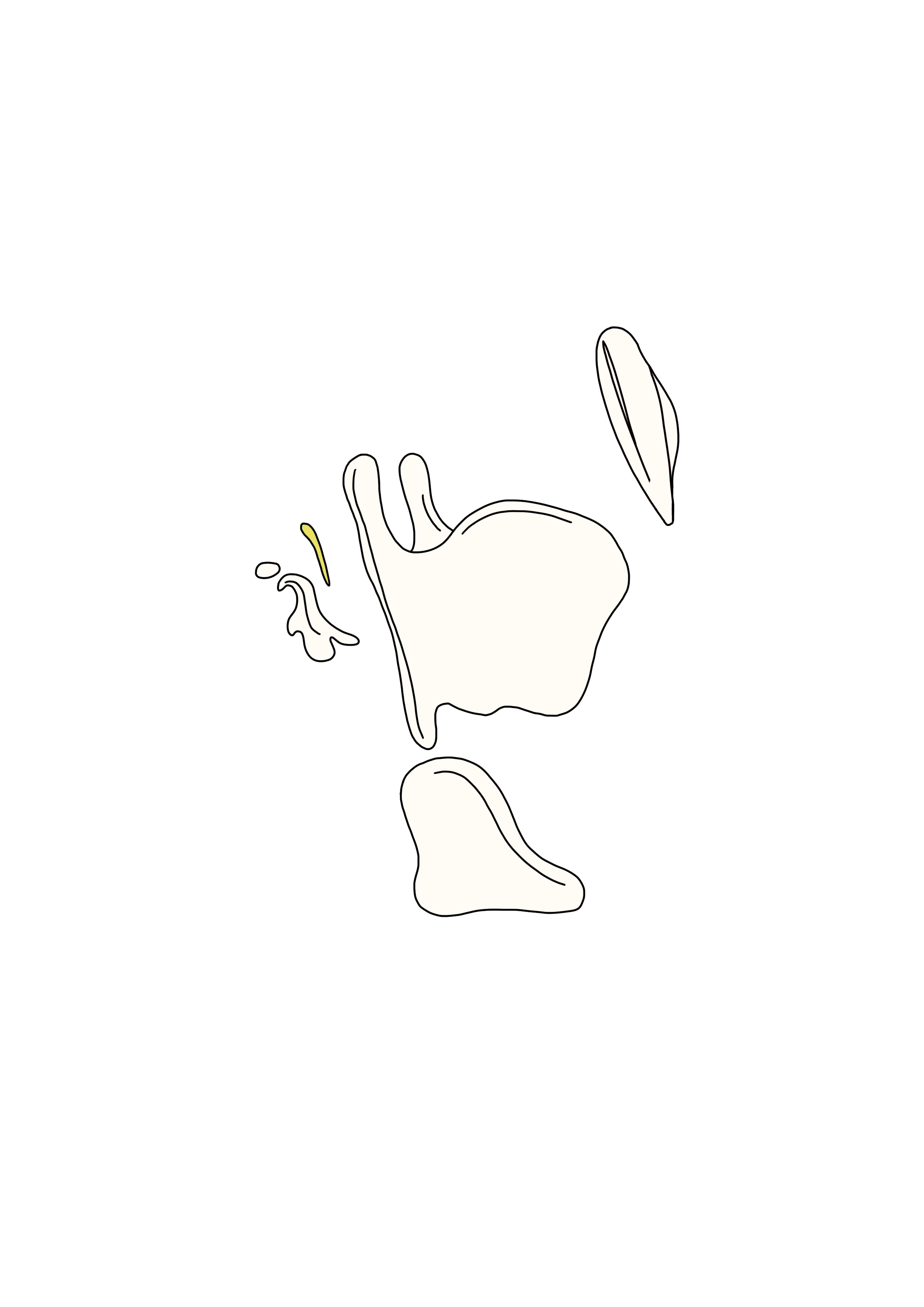
And here they are apart.
There are 2 different types of cartilage referenced below in relation to the larynx:
Hyaline cartilage - primarily type 2 collagen & proteoglycan, glassy, firm and flexible but less elastic, provides a smooth surface for joint movement
Elastic cartilage - contains more elastic fibres, yellowish, flexible, gives structural support whilst also bending
Unpaired Cartilages

The Epiglottic Cartilage
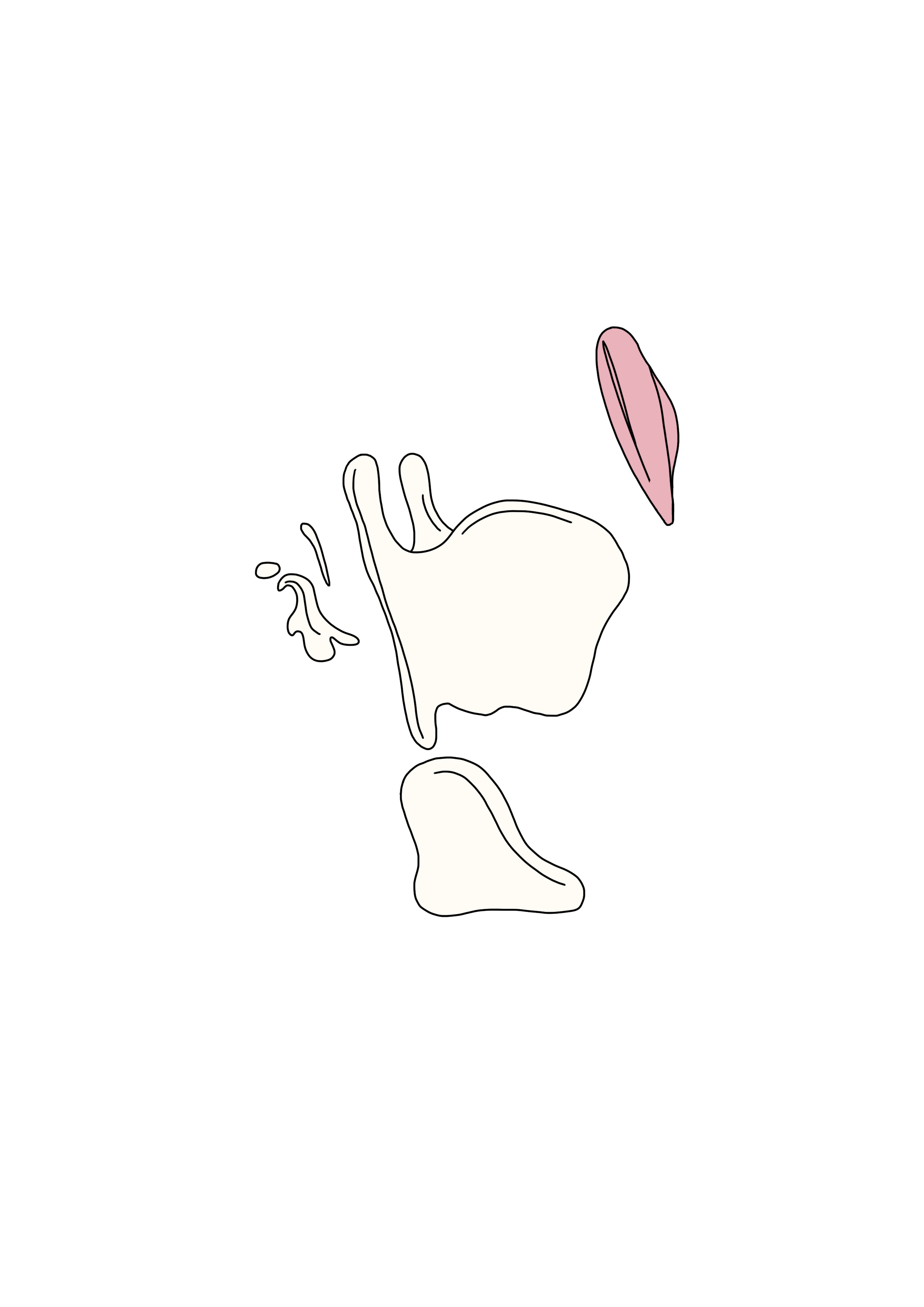
Elastic cartilage
Leaf shaped
Attaches to the hyoid bone via the hyoepiglottic ligament, and attaches to the thyroid cartilage via the thyroepiglottic ligament
Closes the laryngeal inlet during swallowing - this directs food towards the oesophagus rather than the trachea, and protects you from aspirating
The Thryoid Cartilage
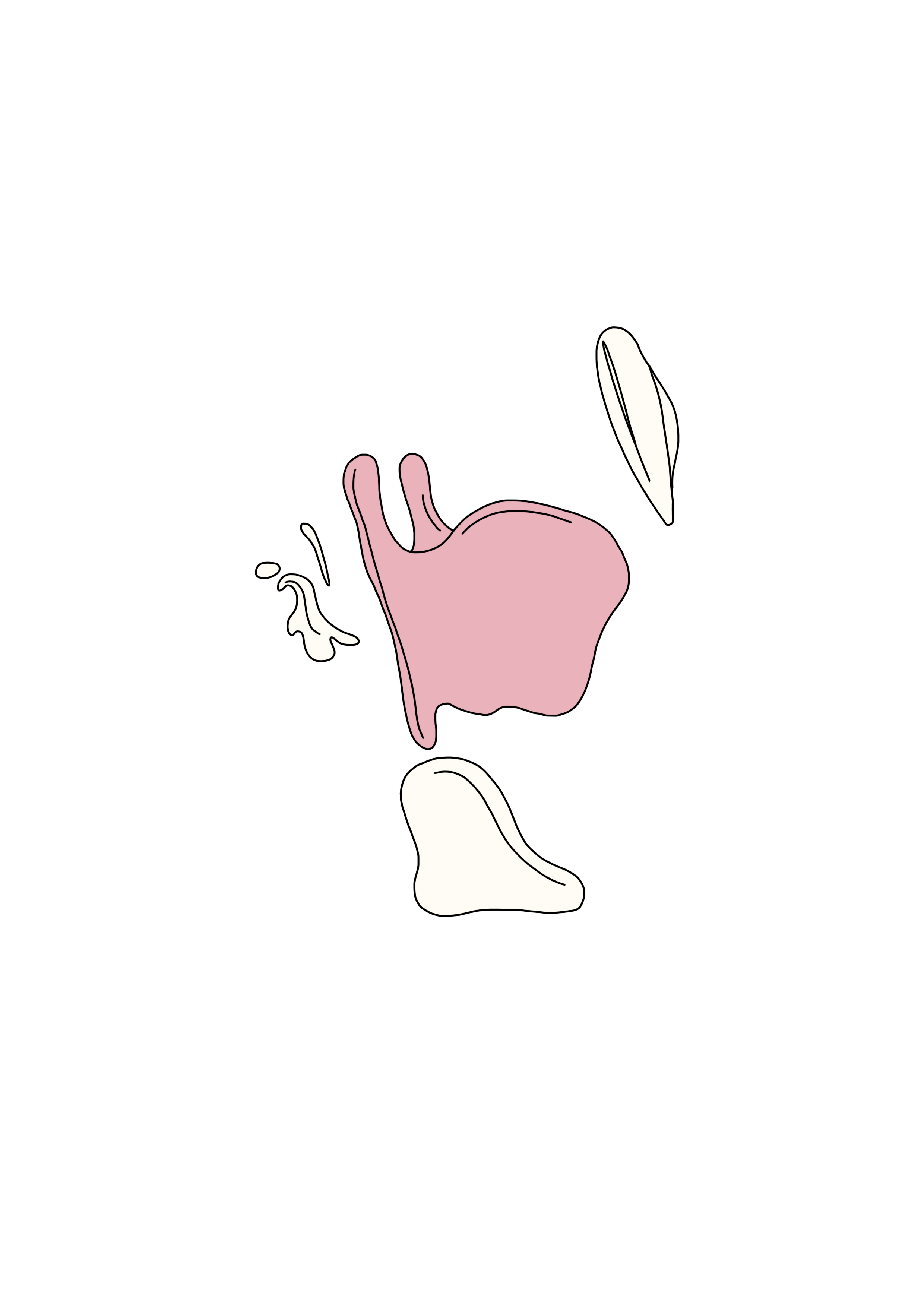
Hyaline cartilage
Consists of two laminae that meet anteriorly at the laryngeal prominence
The posterior borders form the superior and inferior horns (cornua)
The inferior horns articulate with the cricoid cartilage to form cricothyroid joints
The Cricoid Cartilage
Hyaline cartilage
Shaped like a signet ring
Forms a complete ring of cartilage around the airway
Articulates with the thyroid and arytenoid cartilages
Clinical Relevance - Cricoid Pressure
Intubation = tube inserted down the trachea to assist in breathing
‘Cricoid pressure’ may be applied during intubation in patient groups where there is a risk of regurgitated contents coming out of the oesophagus and slipping into the trachea
As the cricoid forms a complete ring, putting to pressure on it will push all the way through and compress the oesophagus
Paired Cartilages

Arytenoid Cartilages

Hyaline cartilage, but contain elastic cartilage at the vocal processes
Pyramidal shape, with a concave base and articular facet superiorly for the corniculate cartilage
It articulates with both the cricoid & corniculate cartilages
They have a muscular process for the attachment of the lateral and posterior cricoarytenoid muscles
And a vocal process for the vocalis muscle, important for the movement of the vocal cords
Corniculate Cartilages
Elastic cartilage
Articulate with the arytenoid cartilage to prolong them posteriorly and medially
Cuneiform Cartilages
Elastic cartilage
Lie within the aryepiglottic folds
Oop - that’s 5 minutes up!
For your next 5 minute session, we suggest…
Laryngeal Muscles
Laryngeal Ligaments & Folds
Laryngeal Vasculature & Innervation
All available in Tamra’s Notes! See you next time x
All original illustrations featured in this blog post are the exclusive intellectual property of the author/creator, Dr Tamra Ariane Nathan. They are protected by copyright laws and must not be copied, reproduced, distributed, altered, or used in any form without the express written permission of the author. Unauthorised use of these illustrations may constitute a violation of intellectual property rights and could result in legal action. If you wish to request permission for usage, please contact the author directly.